Every tool we use is chosen for one reason: to make implant surgery more precise, more predictable, and more comfortable for the patient. From CBCT-guided planning to chairside zirconia milling, the Advani Implant Institute operates as a fully digital, end-to-end surgical center.
Most implant practices outsource portions of the workflow — scans to an outside lab, restorations to a milling center, planning to a third-party radiology service. The Advani Implant Institute does all of it in-house. CBCT imaging, surgical planning, intraoral scanning, model printing, prosthesis milling, and same-day temporary fabrication happen under one roof, on one team, with one surgeon overseeing every step.
This isn't about technology for its own sake. It's about shorter timelines, fewer hand-offs, more predictable results, and the ability to plan, execute, and deliver a same-day full-arch reconstruction without ever sending the patient elsewhere.
For full-arch implant cases, the single biggest source of prosthetic failure is inaccurate impressions. Traditional methods — open-tray analog impressions, even high-end intraoral scans — can introduce small errors that, multiplied across six implants, produce ill-fitting bridges, prosthetic strain, and screw loosening over time.
Photogrammetry solves this with optics. The MicronMapper system uses precision-calibrated cameras to triangulate the exact 3D position of every implant from coded markers attached to each one. The result is a sub-micron map of the implant positions — accuracy that no impression material can match.
The conventional dental impression — a tray of putty held in the mouth for several minutes — is uncomfortable, gag-reflex-triggering, and prone to distortion. The institute uses two of the most advanced intraoral scanners on the market: the 3Shape TRIOS 5 and the Shining 3D Aoralscan Elf.
Both produce high-resolution 3D models of the teeth and arches in under three minutes of scanning, with full color, gingival detail, and bite registration. The scans feed directly into our planning software, milling units, and 3D printers — no shipping, no plaster models, no waiting on a lab.
The final restoration on most of our cases — single crowns, multi-unit bridges, full-arch zirconia prostheses — is milled in-house on a 5-axis CNC milling unit. That means Dr. Advani designs and approves every prosthesis personally, and the patient never waits weeks for an outside lab to deliver the work.
Zirconia is the strongest aesthetic restorative material available. It withstands the bite forces of full-arch function, resists fracture, and — milled at this level of precision — looks remarkably natural. For full-arch cases especially, in-house milling means same-day delivery is possible, and any adjustments happen in the same building, on the same day.
Before every surgical case, we 3D print a physical model of the patient's actual jaw — derived from their CBCT scan and intraoral records. That model lets the surgical team rehearse the case in advance: drill paths, implant angles, soft-tissue management, prosthesis fit. By the time the patient is in the chair, the case has already been done once, on plastic.
The institute runs multiple 3D printers, in parallel, for two main purposes: surgical models for case planning, and same-day temporary bridges that the patient leaves with on the day of surgery. The temporary bridge is designed in the morning, printed during the procedure, and delivered before the patient walks out.
Every implant case starts with a cone-beam CT scan — a 3D radiograph that maps the bone, sinuses, nerves, and existing teeth in volumetric detail. From the scan, every implant is planned virtually before the patient sits in the chair: which implant in which position, at what angle, to what depth, avoiding which anatomy.
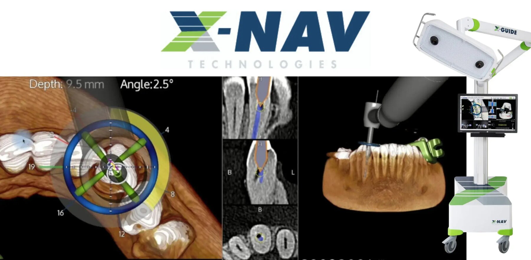
Every case is planned in the two most advanced implant planning platforms available — Blue Sky Plan for full-case virtual planning and prosthetic-driven design, and X-Guide for surgical trajectory planning and navigation setup. Both platforms use AI-assisted anatomical segmentation and trajectory checking. Two independent plans, one converged workflow. If the plans agree, we operate. If they diverge, we investigate why before the drill ever touches bone.
Most implant journeys involve three or four offices: an imaging center for the CBCT, a periodontist or oral surgeon for the surgery, a general dentist for the restoration, and a separate laboratory milling the prosthesis. Every hand-off introduces error and delay.
At the institute, the imaging, planning, surgery, prosthesis design, milling, and delivery all happen in the same building, on the same digital workflow, overseen by Dr. Advani from start to finish. One team, one plan, one continuous experience for the patient.
Six interlocking technologies, applied to every case.
Real-time surgical GPS — implants placed exactly where planned, verified live during surgery.
Sub-micron implant position mapping for passive-fit full-arch prosthetics.
High-resolution intraoral scanning — no impression trays, no waiting on a lab.
Crowns, bridges, and full-arch prostheses milled in-house, designed by Dr. Advani.
Surgical models for case rehearsal; same-day printed temporary bridges.
In-office 3D imaging plus every case planned twice — in Blue Sky Plan and X-Guide — before any drill touches bone.
Every system in our digital stack exists to make implant treatment more precise, more predictable, and more comfortable. The result patients notice: shorter timelines, fewer appointments, and same-day teeth that fit.